elvis15767
New member
Mri results.. not sure exactly what they mean though... any inputs would be appreciated
UPPER EXT MRI JOINT W/ C RIGHT:
MR ARTHROGRAM OF THE RIGHT SHOULDER WITH CONTRAST:
COMPARISON: Right shoulder arthrogram dated 5/9/2017..
TECHNIQUE:
After the intra-articular administration of diluted gadolinium into the right glenohumeral joint under fluoroscopic guidance, MR
images of the right shoulder were performed with an MR shoulder arthrogram protocol.
FINDINGS:
ROTATOR CUFF AND ASSOCIATED STRUCTURES:
Evaluation is degraded due to patient body habitus/tissue composition. This results in poor fat saturation.
Rotator Cuff:
There is intermediate signal throughout the rotator cuff. There is no complete or bursal/articular sided partial rotator cuff tear. The
subscapularis constituent of the rotator cuff is intact.
Bursa: There is a small amount of fluid in the subacromial subdeltoid bursa.
Musculature: There is no muscular tear, contusion or atrophy.
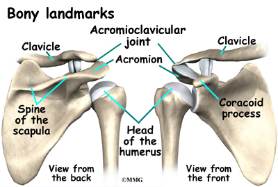
Acromioclavicular joint: There is a type III hooked acromion. There is narrowing of the rotator cuff outlet.. There appears to be mild
superior displacement of the distal clavicle relative to the acromion, demonstrated on the arthrogram images as well. The inferior
acromioclavicular ligament appear disrupted. The superior acromioclavicular ligament appears thickened and edematous but appear
intact. The coracoclavicular ligaments appear normal. There is a subchondral fracture of the distal clavicle. There are erosive changes
at the distal clavicle. There is marrow edema at the distal clavicle. There are areas of cystic change within the distal clavicle. There is
also marrow edema involving the opposing surface of the acromion.
OSSEOUS STRUCTURES:
Bones: There are no other fractures or other regions of abnormal bone marrow signal intensity.
GLENOHUMERAL JOINT:
Cartilage and Bone: No focal hyaline cartilage defects are noted. No Hill-Sach's lesions, or bony Bankart lesions are seen.
Labrum: There are no SLAP or soft tissue Bankart lesions. No paralabral cysts are seen.
OTHER SUPPORT STRUCTURES:
No capsular or ligamentous abnormality is seen. The long head of the biceps tendon is well situated within the bicipital groove without
evidence of tearing or dislocation.
OTHER FINDINGS: None.
IMPRESSION:
1. NORMAL ROTATOR CUFF.
2. NO LABRAL TEARING.
3. SUBCHONDRAL FRACTURE OF THE DISTAL CLAVICLE WITH EROSIVE CHANGES AT THE DISTAL CLAVICLE
AND MARROW EDEMA. FINDINGS REPRESENT DISTAL CLAVICULAR OSTEOLYSIS, LIKELY RELATED TO
REPETITIVE TRAUMA.
4. LOW-GRADE INJURY AT THE ACROMIOCLAVICULAR JOINT, AS ABOVE.
5. TYPE III HOOKED ACROMION WHICH NARROWS THE ROTATOR CUFF OUTLET.
UPPER EXT MRI JOINT W/ C RIGHT:
MR ARTHROGRAM OF THE RIGHT SHOULDER WITH CONTRAST:
COMPARISON: Right shoulder arthrogram dated 5/9/2017..
TECHNIQUE:
After the intra-articular administration of diluted gadolinium into the right glenohumeral joint under fluoroscopic guidance, MR
images of the right shoulder were performed with an MR shoulder arthrogram protocol.
FINDINGS:
ROTATOR CUFF AND ASSOCIATED STRUCTURES:
Evaluation is degraded due to patient body habitus/tissue composition. This results in poor fat saturation.
Rotator Cuff:
There is intermediate signal throughout the rotator cuff. There is no complete or bursal/articular sided partial rotator cuff tear. The
subscapularis constituent of the rotator cuff is intact.
Bursa: There is a small amount of fluid in the subacromial subdeltoid bursa.
Musculature: There is no muscular tear, contusion or atrophy.
Acromioclavicular joint: There is a type III hooked acromion. There is narrowing of the rotator cuff outlet.. There appears to be mild
superior displacement of the distal clavicle relative to the acromion, demonstrated on the arthrogram images as well. The inferior
acromioclavicular ligament appear disrupted. The superior acromioclavicular ligament appears thickened and edematous but appear
intact. The coracoclavicular ligaments appear normal. There is a subchondral fracture of the distal clavicle. There are erosive changes
at the distal clavicle. There is marrow edema at the distal clavicle. There are areas of cystic change within the distal clavicle. There is
also marrow edema involving the opposing surface of the acromion.
OSSEOUS STRUCTURES:
Bones: There are no other fractures or other regions of abnormal bone marrow signal intensity.
GLENOHUMERAL JOINT:
Cartilage and Bone: No focal hyaline cartilage defects are noted. No Hill-Sach's lesions, or bony Bankart lesions are seen.
Labrum: There are no SLAP or soft tissue Bankart lesions. No paralabral cysts are seen.
OTHER SUPPORT STRUCTURES:
No capsular or ligamentous abnormality is seen. The long head of the biceps tendon is well situated within the bicipital groove without
evidence of tearing or dislocation.
OTHER FINDINGS: None.
IMPRESSION:
1. NORMAL ROTATOR CUFF.
2. NO LABRAL TEARING.
3. SUBCHONDRAL FRACTURE OF THE DISTAL CLAVICLE WITH EROSIVE CHANGES AT THE DISTAL CLAVICLE
AND MARROW EDEMA. FINDINGS REPRESENT DISTAL CLAVICULAR OSTEOLYSIS, LIKELY RELATED TO
REPETITIVE TRAUMA.
4. LOW-GRADE INJURY AT THE ACROMIOCLAVICULAR JOINT, AS ABOVE.
5. TYPE III HOOKED ACROMION WHICH NARROWS THE ROTATOR CUFF OUTLET.